By Chinedu Akpa. B. Pharm. Freelance Health Writer and DLHA Volunteer. Medically reviewed by the DLHA Team

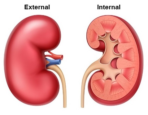
Views of the kidney Image credit: Cleveland Clinic
It is well known that most humans have two kidneys, but did you know that some people are born with an extra kidney? Yes, you heard that correctly. Such people have three kidneys. In medicine, this condition is known as supernumerary (extra or additional) kidney, and it is rare.
Because of the kidney's life-sustaining function, many people have used metaphors like life purifiers, silent workers, chemical factories, the body's balancers, the body's waste thermostat, the hidden guardians, and resilient organs to describe this vital organ. These metaphors describe the kidney and what it does for the body.
It is critical that you understand the structure and functions of the kidney so that when you notice something wrong in your body, you can seek help early.
.jpg) How the Kidney Develops
How the Kidney DevelopsThe kidney begins to develop shortly after the egg is fertilized to form the embryo in week three of pregnancy and the development is completed after 36 weeks of gestation. [1]
From the early phase temporal kidneys (pronephros and mesonephros) to the permanent kidney (metanephros), numerous chemical signals between cells and pathways are established to guarantee the kidney's ultimate formation. A genetic mutation and environmental factors, such as maternal diabetes and exposure to certain antihypertensive medications, can cause problems during the formation of the kidney leading to abnormalities like duplex ureters, horseshoe kidney (kidneys fused in their normal position at their lower pole thereby forming a U shape), absence of a kidney on one side, etc. (See figure 1) [2] Kidney abnormalities may be the cause of clinical complications later in life.
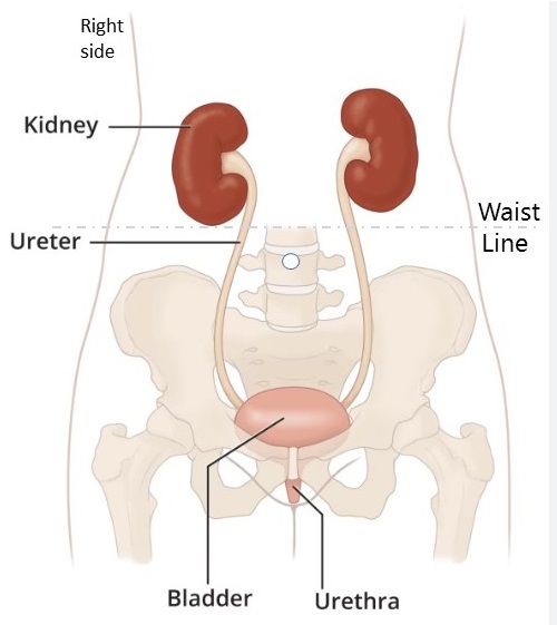
The kidneys are located on each side of the spine, just above the waistline. Due to the liver's location directly above the right kidney, which limits room for further growth, the left kidney is typically larger and located slightly higher than the right (Figure 2). Each kidney is connected to the bladder by the ureter.

Figure 2: Showing location of the kidneys. Image: Adapted from Cleveland Clinic
The kidney is roughly the size of a closed fist, and it’s shaped like a bean. In both males and females, they weigh differently. As would be expected, the kidney weighs higher in men; 150–200 g and 120–135 g in women. Their measurements are 10–12 cm in length, 5-7 cm in width, and 3-5 cm in thickness. [3]
Figure 3: Diagram of the nephron with labeled parts and direction of urine flow.
Nephrons are the kidney's functional units, just like the body's cells are its units of function. Approximately one million nephrons are found in each kidney, and they are constantly working. [4]
Nephrons, despite their small size, are made up of various parts that work together to provide a whole function. These components include the Glomerulus, Bowman’s capsule, Proximal Convoluted Tube, Loop of Henle, Distal Convoluted Tubule, Collecting Duct. The functions of these parts of the nephrons will be discussed later in this article, stay with us!

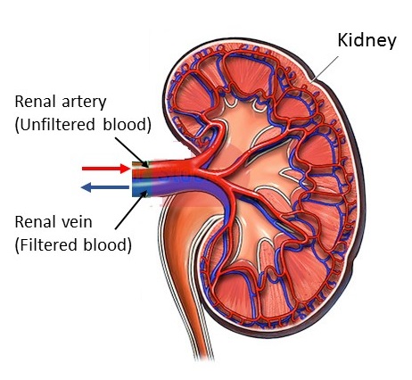
Figure 4: Blood supply of the kidney with direction of flow.
The kidney's blood supply is essential to its operation. The kidney receives one of the highest amounts of blood in the body—roughly 22 percent of the blood pumped out by the heart goes directly to it. [5]
The kidney is assisted in eliminating waste from the body by the pressure created when the heart pumps blood to the kidney. The kidney can be likened to a water purification plant that is fed by the continuous flow of water from a river (the heart's pumping action) in order to function. The plant cannot adequately filter the water if the river flows too slowly. However, if the water flows too quickly, it may overload the plant and harm its fragile machinery. Continuing with the comparison of the water purification plant, the human kidney is able to filter the water and maintain a clean system because the heartbeat regulates the blood flow to it. The renal vein and artery are the two blood vessels that are connected to the kidney.
The only blood vessels that supply unfiltered blood to the kidney are the renal arteries. [6] The red colour of the renal artery in the above diagram (figure 2) indicates that it is delivering oxygen-rich blood straight from the heart to the kidneys. The kidney filters this blood. [5]
They are the only veins that drain blood from the two kidneys. They carry filtered blood away from the kidney to another vessel called the inferior vena cava on its way to the heart. [7]
The blue colour of the renal vein in figure 2, indicates that it is carrying blood that lacks oxygen away from the kidney to the heart.
Published: September 14, 2024
© 2024. Datelinehealth Africa Inc. All rights reserved.
Permission is given to copy, use and share content freely for non-commercial purposes without alteration or modification and subject to source attribution
DATELINEHEALTH AFRICA INC., is a digital publisher for informational and educational purposes and does not offer personal medical care and advice. If you have a medical problem needing routine or emergency attention, call your doctor or local emergency services immediately, or visit the nearest emergency room or the nearest hospital. You should consult your professional healthcare provider before starting any nutrition, diet, exercise, fitness, medical or wellness program mentioned or referenced in the DatelinehealthAfrica website. Click here for more disclaimer notice.